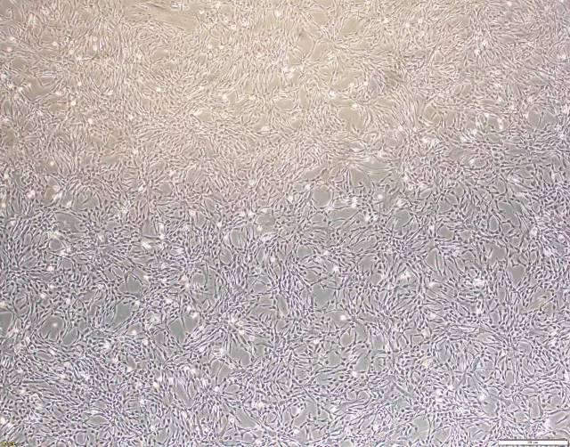
动物试验早就证明间充质干细胞能有效地治疗肝衰竭[1, 2],而且直接用间充质干细胞治疗肝衰竭效果优于用间充质干细胞进行诱导分化后的肝细胞[3]。动物实验的数据和研究的深入,同时伴随着临床研究的发展。
2006年,Terai等用自体骨髓干细胞治疗9名肝硬化患者,发现经过骨髓干细胞治疗后,患者肝功能得到明显改善[4]。这是间充质干细胞治疗肝病的第一篇临床研究报道。在2007年及2011年,两个研究小组均进一步验证了骨髓干细胞改善肝功能的疗效[5, 6]。骨髓干细胞虽然能短期改善肝功能,但长期效果欠佳。而且,骨髓干细胞包含了造血干细胞和间充质干细胞,以及少量的干/祖细胞。因此很难评价骨髓干细胞改善肝功能是通过哪类干细胞起作用的。
2009年瑞典报道了自体骨髓间充质干细胞治疗肝硬化的I-II期临床试验结果,进一步证明了骨髓间充质干细胞治疗能降低终末期肝病评分(End-Stage Liver Disease Score),改善肝功能,降低转氨酶和胆红素,而且没发现不良反应[7]。
2011年中山大学第三附属医学院报道了骨髓间充质干细胞治疗(乙肝)肝衰竭的大样本临床研究,间充质干细胞治疗(单次剂量、肝脏介入治疗)的短期(一个月)效果明显,肝功能改善,MELD评分降低,但是治疗组和对照组的长期生存率(约50%)没有差异,肝癌的发生率也没有差异[8]。遗憾的是,这篇文献并没有提及肝衰竭患者接受骨髓间充质干细胞治疗的细胞数量。一年后的2012年,北京302医院报道了脐带间充质干细胞治疗乙肝肝衰竭(慢加急)的临床研究结果,按照0.5x10⁶/kg的细胞数,每4周治疗一次,治疗3次;和对照组相比,治疗组肝功能改善明显,MELD评分降低,而且长期生存率提高到约70%(对照组为40%)[9]。
这两篇文献报道结果的差异,可能存在至少两方面的影响:①间充质干细胞来源的不同,和②间充质干细胞治疗方案的差异。有研究发现脐带来源的间充质干细胞分泌的肝细胞生长因子(hepatic growth factor, HGF)是骨髓来源间充质干细胞的30倍[10]。而HGF是最重要的促进肝细胞增殖和肝脏修复的蛋白因子[11, 12]。目前,依然需要深入的大样本的临床研究,优化治疗方案,以期达到更佳的治疗效果和更高的长期生存率。
肝衰竭的病理基础在于肝细胞的大量坏死,其治疗效果取决于肝脏的肝细胞是否及时再生到一定的数量,以维持肝脏的各种基本功能(合成、代谢、解毒等)。卵圆细胞被认为是肝细胞的前体/干细胞细胞,当肝脏轻微损伤时肝脏可通过自身的肝细胞增殖修复,如果肝脏严重受损致使肝细胞大部分坏死,肝内前体/干细胞——肝卵圆细胞便被激活并分化生成肝细胞和胆管细胞等参与肝修复[13]。SDF-1α是卵圆细胞重要的激活因子,间充质干细胞通过分泌细胞因子(SDF-1α)激活卵圆细胞、促进肝细胞再生作用[14, 15]。当然,除了HGF和SDF-1α外,可能还存在其他的未知的细胞因子发挥了修复肝脏的作用,这均需要深入的科学研究。
关于间充质干细胞治疗肝衰的机制,目前比较一致的看法就是间充质干细胞分泌各种因子,抑制了免疫反应和炎症反应,促进的肝脏组织器官的修复和内源性肝脏干细胞的增殖,促进肝细胞的再生,从而达到了治疗效果,而和间充质干细胞的分化功能无明显关系[1, 16-18]。
参考文献:
[1]Kuo T. K., Hung S. P.,Chuang C. H., et al. Stem cell therapy for liver disease: parameters governingthe success of using bone marrow mesenchymal stem cells. Gastroenterology2008;134(7):2111-2121, 2121 e2111-2113.
[2]Banas A., Teratani T., Yamamoto Y., et al. Rapidhepatic fate specification of adipose-derived stem cells and their therapeuticpotential for liver failure. J Gastroenterol Hepatol 2009;24(1):70-77.
[3]Wang H., Zhao T., Xu F., et al. How important isdifferentiation in the therapeutic effect of mesenchymal stromal cells in liverdisease? Cytotherapy 2014;16(3):309-318.
[4]Terai S., Ishikawa T., Omori K., et al. Improvedliver function in patients with liver cirrhosis after autologous bone marrowcell infusion therapy. Stem Cells 2006;24(10):2292-2298.
[5]Lyra A. C., Soares M. B., Da Silva L. F., et al.Feasibility and safety of autologous bone marrow mononuclear celltransplantation in patients with advanced chronic liver disease. World JGastroenterol 2007;13(7):1067-1073.
[6]Saito T., Okumoto K., Haga H., et al. Potentialtherapeutic application of intravenous autologous bone marrow infusion inpatients with alcoholic liver cirrhosis. Stem Cells Dev 2011;20(9):1503-1510.
[7]Kharaziha P., Hellstrom P. M., Noorinayer B., etal. Improvement of liver function in liver cirrhosis patients after autologousmesenchymal stem cell injection: a phase I-II clinical trial. Eur JGastroenterol Hepatol 2009;21(10):1199-1205.
[8]Peng L., Xie D. Y., Lin B. L., et al. Autologousbone marrow mesenchymal stem cell transplantation in liver failure patientscaused by hepatitis B: short-term and long-term outcomes. Hepatology2011;54(3):820-828.
[9]Shi M., Zhang Z., Xu R., et al. Human mesenchymalstem cell transfusion is safe and improves liver function in acute-on-chronicliver failure patients. Stem Cells Transl Med 2012;1(10):725-731.
[10]Friedman R., Betancur M., Boissel L., et al.Umbilical cord mesenchymal stem cells: adjuvants for human celltransplantation. Biol Blood Marrow Transplant 2007;13(12):1477-1486.
[11]Fujiwara K., Nagoshi S., Ohno A., et al.Stimulation of liver growth by exogenous human hepatocyte growth factor innormal and partially hepatectomized rats. Hepatology 1993;18(6):1443-1449.
[12]Matsumoto K., Nakamura T. Hepatocyte growthfactor: molecular structure and implications for a central role in liverregeneration. J Gastroenterol Hepatol 1991;6(5):509-519.
[13]Petersen B. E., Bowen W. C., Patrene K. D., etal. Bone marrow as a potential source of hepatic oval cells. Science1999;284(5417):1168-1170.
[14]Zheng D., Oh S. H., Jung Y., et al. Oval cellresponse in 2-acetylaminofluorene/partial hepatectomy rat is attenuated byshort interfering RNA targeted to stromal cell-derived factor-1. Am J Pathol2006;169(6):2066-2074.
[15]Hatch H. M., Zheng D., Jorgensen M. L., et al.SDF-1alpha/CXCR4: a mechanism for hepatic oval cell activation and bone marrowstem cell recruitment to the injured liver of rats. Cloning Stem Cells2002;4(4):339-351.
[16]Parekkadan B., Van Poll D., Suganuma K., et al.Mesenchymal stem cell-derived molecules reverse fulminant hepatic failure. PLoSOne 2007;2(9):e941.
[17]Van Poll D., Parekkadan B., Cho C. H., et al.Mesenchymal stem cell-derived molecules directly modulate hepatocellular deathand regeneration in vitro and in vivo. Hepatology 2008;47(5):1634-1643.
[18]Chen Y. X., Zeng Z. C., Sun J., et al.Mesenchymal stem cell-conditioned medium prevents radiation-induced liverinjury by inhibiting inflammation and protecting sinusoidal endothelial cells.J Radiat Res 2015;56(4):700-708.
本文转自:湖北省生命源干细胞(微信号hbsmy2010)